


Acupuncture points have a higher density of micro-vessels and contain a large amount of involuted microvascular structures. The non-acupuncture points did not exhibit these properties. The researchers note that the state-of-the-art CT imaging techniques used in this study allow for improved three-dimensional (3D) imaging of a large field of view without artifacts. This greatly improves imaging of soft tissue and allowed the researchers to make this important discovery.
The acupuncture points ST36 (Zusanli) and ST37 (Shangjuxu) were shown to have very distinct structural differences than surrounding areas. At the acupuncture points, microvascular densities with bifurcations “can be clearly seen around thick blood vessels” but non-acupuncture point areas showed few thick blood vessels and none showed fine, high density structures. The acupuncture points contained fine structures with more large blood vessels that are several dozen micrometers in size plus beds of high density vascularization of vessels 15-50 micrometers in size. This structure was not found in non-acupuncture point areas.
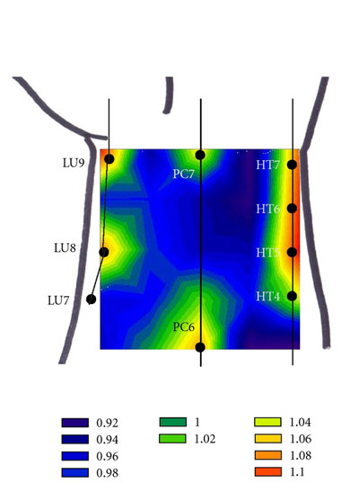
In another interesting study, researchers used an amperometric oxygen microsensor to detect partial oxygen pressure variations at different locations on the anterior aspect of the wrist. The researchers concluded that partial oxygen pressure is significantly higher at acupuncture points. Below are images from the study measuring the increase of partial oxygen pressure combined with an overlay of the local acupuncture point locations. The images map the Lung, Pericardium and Heart channels and their associated local points. Acupuncture points P7 and P6 clearly show high oxygen pressure levels as do the other acupuncture points in the region.
These measurements are not needled points but are natural resting states of acupuncture points absent of stimulation. A truly unique finding, acupuncture points exhibit special oxygen characteristics. Acupuncture points and acupuncture channels are scientifically measurable phenomena in repeated experiments.
Nice post! acupuncture is a very good method to do that. She 'reads' our wrist and feels how the energy (qi) flows in our bodies, where blockages are located, if you have a virus, etc. It's an ancient Chinese medical technique and it works fine for me. Thanks for sharing information.
ReplyDeleteAcupuncture in Chicago